

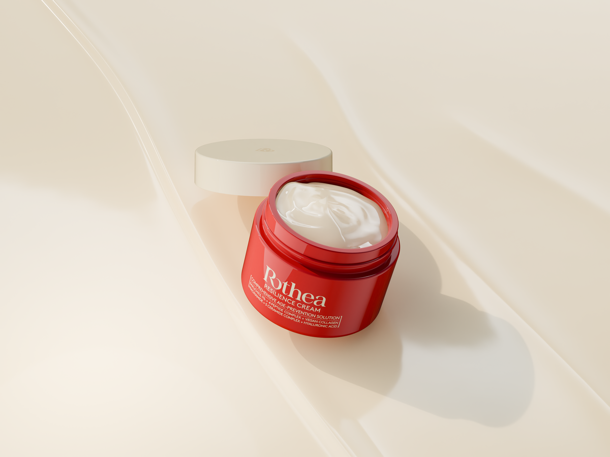
Managing hyperpigmentation while simultaneously targeting the structural signs of aging is one of the most complex balancing acts in modern dermatology. Consumers dealing with melasma, post-inflammatory hyperpigmentation (PIH), and age-associated dark spots face a frustrating paradox: the very ingredients traditionally heralded for accelerating youthful skin (such as high-strength retinoids, aggressive alpha-hydroxy acids [AHAs], and intense chemical peels) frequently trigger an inflammatory response that exacerbates pigment production.
For skin types prone to hyperactive melanogenesis, traditional anti-aging protocols represent a high-risk gamble. The path to achieving a smooth, even, and youthful complexion does not require forcing the skin into a state of chronic crisis. Rather, it requires a sophisticated, non-inflammatory strategy that regulates melanin synthesis pathways while simultaneously reinforcing barrier integrity and boosting collagen production.
This comprehensive guide explores the cellular mechanisms behind pigment rebound, details why aggressive treatments fail pigment-prone skin, and introduces bakuchiol as a clinically proven, botanically derived alternative capable of safely fading hyperpigmentation without skin irritation.
Chapter 1: The Cellular Matrix of Hyperpigmentation: Understanding Melasma and PIH
To successfully address hyperpigmentation, one must first understand the biological machinery driving it. The skin’s pigmentary system is a highly sensitive defense mechanism designed to protect cellular DNA from stressors such as ultraviolet (UV) radiation, environmental pollution, and physical trauma.
The Role of the Melanocyte
Melanocytes are specialized cells located in the basal layer of the epidermis. They contain organelles called melanosomes, within which the amino acid tyrosine is converted into melanin via the rate-limiting enzyme tyrosinase. Once synthesized, these melanin-rich melanosomes are transported via dendritic arms to neighboring keratinocytes, where they form a protective, umbrella-like cap over the cell nuclei to absorb harmful radiation.
Melasma vs. Post-Inflammatory Hyperpigmentation (PIH)
While both conditions manifest as excess pigment, their underlying etiologies differ significantly:
-
Melasma: A chronic, multifactorial disorder characterized by symmetrical macules and patches of hyperpigmentation, primarily on the face (cheeks, upper lip, forehead, and chin). It is uniquely sensitive to hormonal fluctuations (estrogen and progesterone), heat (infrared radiation), vascular endothelial growth factor (VEGF), and UV light. Melasma melanocytes are chronically hyperactive and biologically distinct from normal melanocytes.
-
Post-Inflammatory Hyperpigmentation (PIH): An acquired hypermelanosis that occurs following cutaneous injury or inflammation, such as acne, eczema, or aggressive cosmetic procedures. Any disruption to the epidermal basement membrane triggers a surge in inflammatory mediators (including prostaglandins, leukotrienes, and interleukins) which directly stimulate melanocytes to overproduce pigment.
Chapter 2: The Hyperpigmentation Trap
The fundamental flaw in conventional anti-aging philosophies for pigment-prone skin is the reliance on controlled trauma. The theory states that by injuring the skin superficially, you kickstart the body’s natural wound-healing response, resulting in fresh collagen synthesis and rapid cellular turnover. While robust skin can tolerate this cycle, sensitive and hyperpigmented skin types frequently experience a severe pigmentary backlash known as the rebound effect.
The Destructive Path of Strong Chemical Peels and High-Percentage Acids
High-concentration glycolic acid peels, Jessner’s solutions, and unbuffered trichloroacetic acid (TCA) treatments work by rapidly cleaving the desmosomes holding dead skin cells together. When applied incorrectly or too aggressively, they cause:
-
Acute Stratum Corneum Desquamation: Stripping the outermost protective layer eliminates the skin’s physical shield against the environment.
-
Trans-Epidermal Water Loss (TEWL): Moisture quickly evaporates from the unprotected lower layers of the skin, inducing structural cellular stress.
-
Pro-Inflammatory Cytokine Cascade: The sudden cellular trauma releases alarm signals like tumor necrosis factor-alpha (TNF-α) and interleukin-1 (IL-1). These cytokines migrate directly to the basal layer, activating the melanocyte-stimulating hormone (α-MSH) receptor. The melanocytes interpret this as a severe threat and flood the surrounding tissue with emergency melanin, creating darker, deeper, and more stubborn hyperpigmentation than before.
The Problem with High-Concentration Retinoids
Prescription-strength retinoids (such as tretinoin) and high-percentage over-the-counter retinol are excellent tools for upregulating collagen genes and normalizing cellular turnover. However, the initial phase of retinoid usage often introduces retinoid dermatitis. Symptoms include severe erythema (redness), flaking, burning, and pruritus (itching).
For a melasma patient, this localized redness is a precursor to pigment deposition. If the skin barrier remains in a state of chronic inflammation for weeks at a time, the newly synthesized skin cells will be heavily populated with excess melanosomes, rendering the anti-aging effort completely counterproductive.
Chapter 3: The Bakuchiol Breakthrough & Non-Inflammatory Anti-Aging
To bypass the inflammatory pitfalls of standard chemical peels and retinoids, dermatology researchers have sought out ingredients capable of delivering equivalent cellular benefits without altering the skin's tolerance threshold. This search led to the identification of bakuchiol.
| Characteristic | Traditional Retinoids | Bakuchiol |
|---|---|---|
| Source | Synthetic or Animal-Derived | Psoralea corylifolia plant |
| Mechanism of Action | Binds to RAR/RXR receptors | Upregulates similar gene pathways |
| Irritation Potential | High (Retinoid Dermatitis) | Extremely Low |
| Photostability | Unstable (Degrades in sunlight) | Highly Stable (Photoprotective) |
| Melanin Regulation | Indirect (Via rapid turnover) | Direct (Tyrosinase inhibition) |
| Barrier Impact | Initially disruptive | Reinforces & soothes |
What is Bakuchiol?
Bakuchiol is a pure, monoterpene phenol isolated primarily from the seeds and leaves of the Psoralea corylifolia (babchi) plant, a staple of Ayurvedic and traditional Chinese medicine. While structurally distinct from vitamin A derivatives, genomic expression profiling reveals that bakuchiol acts as a functional analogue to retinol. It utilizes the same cellular machinery and upregulates similar genetic pathways in human skin fibroblasts without inducing the irritant pathways that lead to redness and barrier breakdown.
The Science of Gentle Melanin Regulation
Bakuchiol alters the hyperpigmentation landscape by directly intervening at multiple points within the melanogenesis pathway, avoiding the collateral damage caused by harsh exfoliants.
Downregulation of Tyrosinase Activity
Bakuchiol functions as a direct inhibitor of the tyrosinase enzyme. By binding to or interfering with the operational mechanics of tyrosinase, it reduces the conversion rate of tyrosine into L-DOPA and subsequent dopaquinone. Since this inhibition occurs quietly at the enzyme level, the melanocyte does not experience structural trauma or distress, thereby avoiding an inflammatory counter-response.
Stabilization of Cellular Turnover
Unlike chemical peels that physically rip away the stratum corneum, bakuchiol upregulates the genes responsible for normal cellular differentiation and desquamation. It gently guides the skin back to a healthy 28-day turnover cycle. This orderly, non-violent acceleration of cellular renewal allows existing pools of accumulated melanin to clear out of the epidermis systematically, ensuring that newly arrived keratinocytes are evenly pigmented.
Interruption of the Oxidative Stress Cascade
UV radiation and atmospheric pollution generate reactive oxygen species (ROS) within the skin. These free radicals lipid-peroxidize the cellular membranes, signaling the body to produce inflammatory mediators that stimulate melanocytes.
Bakuchiol is a potent lipid-soluble antioxidant. By scavenging these free radicals before they can interact with the cell membrane, it halts the oxidative signal before it can reach the nucleus of the melanocyte.
Maintenance of the Epidermal Barrier
Bakuchiol upregulates the synthesis of critical extracellular matrix components, including collagen types I, III, and IV, as well as aquaporin-3 (a water and glycerol channel protein that maintains epidermal hydration). A robust, well-hydrated stratum corneum keeps environmental irritants out and minimizes the internal cellular friction that provokes inflammatory hyperpigmentation.
Chapter 4: Designing a Synergistic, Melasma-Safe Routine
While bakuchiol is an exceptional standalone ingredient for addressing both aging and hyperpigmentation, a truly comprehensive approach utilizes multiple non-irritating actives. Each ingredient should target a distinct phase of the melanin pathway, creating a multi-layered defense system.
| Target Mechanism | Active Ingredient Solving It |
|---|---|
| 1. Block Inflammatory Signals | Tranexamic Acid |
| 2. Inhibit Tyrosinase Activity | Bakuchiol / Kojic Acid |
| 3. Halt Melanosome Transfer | Niacinamide |
| 4. Prevent UV / Heat Triggering | Zinc Oxide (Mineral SPF) |
The Perfect Pairs: Formulating with Synergistic Brighteners
Niacinamide (Vitamin B3): The Transfer Blocker
Even when tyrosinase activity is mitigated, some melanin will inevitably be produced. This is where niacinamide steps in. Niacinamide specifically inhibits the transfer of melanosomes from the dendritic arms of the melanocytes into the surrounding keratinocytes.
When paired with bakuchiol, you establish a two-zone defense: bakuchiol minimizes the production of the pigment, and niacinamide blocks whatever remains from reaching the visible surface of the skin. Furthermore, niacinamide stimulates ceramides, strengthening the skin barrier against external stressors.
Tranexamic Acid: The Inflammatory Switch
Tranexamic acid is a synthetic lysine analogue that has revolutionized the treatment of melasma. It works by inhibiting the activation of plasminogen to plasmin. In the skin, UV radiation induces keratinocytes to secrete plasminogen activator, which increases arachidonic acid and prostaglandin production. These molecules are powerful stimulators of melanogenesis.
By applying tranexamic acid, you turn off this inflammatory switch, ensuring that the melanocytes remain in a calm, unprovoked state while bakuchiol works on structural collagen restructuring.
Kojic Acid and Azelaic Acid: Selective Tyrosinase Control
For persistent dark spots, combining bakuchiol with mild, non-stripping tyrosinase inhibitors like azelaic acid or kojic acid provides amplified results. Azelaic acid is particularly beneficial because it has a selective affinity for hyperactive melanocytes, leaving normal, healthy pigmented skin cells largely unaffected. It also works as a mild anti-inflammatory agent, making it safe for rosacea-prone skin types that frequently struggle with concurrent melasma.
Step-by-Step Blueprint for a Melasma-Safe Routine
To minimize irritation and maximize anti-aging efficacy, structure your morning and evening routines around barrier protection, pigment regulation, and environmental shielding.
(0.5% - 1.0%)
The Morning (AM) Protocol: Defend and Stabilize
-
Step 1: Gentle Hydrating Cleanser: Use a non-foaming, sulfate-free cleanser that leaves the natural lipid barrier completely intact. Avoid hot water, which induces vasodilation and heat-induced melasma flare-ups; use tepid water instead.
-
Step 2: Pigment-Stabilizing Serum: Apply a water-based serum containing 2-5% Niacinamide combined with 2-3% Tranexamic Acid. This preps the skin to resist daylight inflammatory triggers.
-
Step 3: Lightweight Ceramides Moisturizer: Apply a barrier-replenishing lotion rich in essential fatty acids and phytosterols to seal in moisture and prevent trans-epidermal water loss.
-
Step 4: Broad-Spectrum Mineral Sunscreen (SPF 50+): Formulate strictly with non-nano Zinc Oxide and Titanium Dioxide. Mineral sunscreens sit on top of the skin to reflect UV rays, whereas chemical sunscreens absorb UV rays and convert them into heat energy; a process that can trigger underlying melasma. Ensure the formula contains Iron Oxides, which provide crucial protection against visible blue light from digital screens and daylight, a proven catalyst for deeper pigmentation.
The Evening (PM) Protocol: Rebuild and Correct
-
Step 1: Double Cleansing: Begin with an emollient cleansing oil or balm to dissolve sunscreen, airborne particulate matter, and sebum without stripping the skin. Follow with your AM gentle hydrating cleanser to clear away remaining impurities.
-
Step 2: Bakuchiol Treatment Serum: Apply a dedicated serum containing a clinically validated concentration of pure bakuchiol (typically between 0.5% and 1.0%). Press the serum gently into the face, neck, and décolletage, avoiding aggressive rubbing.
-
Step 3: Deep Recovery Barrier Cream: Finish with a richer cream infused with squalane, hyaluronic acid, and allantoin. This seals in the bakuchiol and supports nocturnal cellular repair, allowing the skin to rest in an un-inflamed, highly nourished state.
Chapter 5: Bakuchiol vs. Retinol
To appreciate bakuchiol's role as a primary treatment for sensitive, pigment-prone skin, look at the landmark clinical evidence validating its real-world performance against traditional anti-aging standards.
A definitive 12-week randomized, double-blind clinical study published in the British Journal of Dermatology directly compared 0.5% bakuchiol cream against 0.5% retinol cream in human subjects over a 12-week period. The trial measured significant parameters including wrinkle surface area, hyperpigmentation intensity, and skin tolerability.
| Measured Parameter | Study Results & Outcomes |
|---|---|
| Wrinkle Reduction | Equal, statistically identical efficacy |
| Hyperpigmentation Clearance | Significant clearance in both groups |
| Skin Scaling & Flaking | High in Retinol / Minimal in Bakuchiol |
| Stinging & Burning Sensations | Frequent with Retinol / Rare with Bakuchiol |
Wrinkle and Fine Line Reduction
The objective cutaneous imaging analysis revealed that both the bakuchiol and retinol cohorts experienced a statistically identical, significant reduction in fine lines and wrinkle surface area. This occurred due to both ingredients' ability to stimulate Type I collagen synthesis via the upregulation of specific target genes in dermal fibroblasts.
Hyperpigmentation Clearance
Both groups demonstrated a notable decrease in hyperpigmented intensity and area at the 12-week mark. Retinol achieved this primarily via rapid epidermal scaling, while bakuchiol achieved it via direct tyrosinase inhibition and controlled cellular turnover.
Tolerability Profiles
This is where the paths diverged. The retinol users reported significantly higher instances of:
-
Skin scaling, flaking, and structural peeling.
-
Severe stinging, burning, and localized pruritus.
-
Persistent erythema (redness).
In contrast, the bakuchiol users experienced minimal to no adverse side effects, demonstrating that a patient can achieve identical pigment clearance and structural wrinkle reversal without enduring the inflammatory side effects that cause melasma to rebound.
Chapter 6: Managing Melasma from the Inside Out
Dermatological topicals form only one pillar of an effective melasma-safe strategy. Since hyperactive melanocytes respond directly to internal and external stimuli, lifestyle adjustments are required to achieve and maintain long-term results.
The Invisible Enemy: Heat and Infrared Radiation (IR)
Many individuals diligently apply sunscreen but notice their melasma flaring up after cooking over a hot stove, taking a hot yoga class, sitting in a sauna, or spending time in an overheated vehicle.
This occurs because heat induces immediate vasodilation (widening of blood vessels) and local cutaneous inflammation. The increased blood flow elevates the expression of vascular endothelial growth factor (VEGF), which directly communicates with basal melanocytes, prompting them to synthesize melanin.
-
Mitigation Strategy: Keep a cooling facial mist or a portable fan nearby during exercise or cooking. Apply a cold compress to the skin immediately after heat exposure to rapidly lower skin temperature and halt the vascular inflammatory response.
Dietary Influences on Cutaneous Inflammation
Chronic systemic inflammation lowers the threshold at which melanocytes react to external stressors. High-glycemic diets rich in refined sugars and processed carbohydrates cause rapid spikes in blood glucose, triggering the release of insulin and insulin-like growth factor 1 (IGF-1). High levels of IGF-1 stimulate cellular inflammatory pathways that can aggravate both acne-driven PIH and hormonal melasma.
-
Dietary Adjustments: Focus on an anti-inflammatory diet rich in polyphenols, omega-3 fatty acids (found in wild-caught salmon and flaxseeds), and cruciferous vegetables like broccoli and Brussels sprouts, which contain indole-3-carbinol to support healthy estrogen metabolism.
Stress, Cortisol, and Hormonal Fluctuations
Emotional and physical stress stimulates the hypothalamic-pituitary-adrenal (HPA) axis, resulting in a systemic surge of cortisol and adrenocorticotropic hormone (ACTH). ACTH shares structural similarities with α-MSH (melanocyte-stimulating hormone). Elevated stress levels can directly bind to and stimulate melanocyte receptors, causing a sudden darkening of facial pigmentation.
-
Stress Management: Incorporate daily mindfulness exercises, sleep support protocols, and regular moderate exercise to normalize systemic cortisol outputs and stabilize pigment production.
Chapter 7: Troubleshooting and Expectation Management
When pivoting from an aggressive anti-aging approach to a gentle, non-inflammatory bakuchiol-based routine, clear expectations and careful monitoring are key.
| ⏰ 2 to 4 Weeks | Improved Barrier & Hydration |
| ⏰ 6 to 8 Weeks | Visible Smoothing & Brightness |
| ⏰ 12+ Weeks | Deep Pigment & Fine Line Loss |
Understanding the Timeline of Gentle Correction
Since bakuchiol works with your skin's natural rhythm rather than forcing it through trauma, changes take time. Hyperpigmentation did not form overnight, and fading it safely requires persistence.
-
Weeks 2-4: The skin barrier strengthens. You should experience decreased redness, minimized flaking, and improved overall skin hydration.
-
Weeks 6-8: Epidermal cellular turnover begins to normalize. The complexion will look brighter, and superficial post-inflammatory dark spots will start to soften at their edges.
-
Weeks 12 and Beyond: Deep-seated melasma patches show visible reduction. Fine lines and structural skin laxity improve as newly synthesized collagen reaches the upper dermal layers.
How to Spot and Address Hidden Inflammation
Even mild skincare routines can cause issues if your skin is extremely reactive. Monitor your skin daily for signs of low-grade inflammation:
-
Micro-Flaking: Fine, powder-like peeling around the corners of the mouth or nose indicates that your skin barrier is struggling to keep up with cellular turnover. Decrease usage frequency or increase your barrier cream application.
-
Transient Erythema: If your skin displays a pink or reddish flush for more than ten minutes post-skincare application, your barrier may be compromised. Temporarily pause all active serums and focus exclusively on a simple cleanser, plain moisturizer, and mineral sunscreen until the redness resolves.
Conclusion: Embracing a Preservation First Philosophy
Fading stubborn hyperpigmentation and reversing structural skin aging does not require aggressive, barrier-stripping treatments. For sensitive, pigment-prone skin, treatments like harsh chemical peels and aggressive retinoids often trigger a reactive rebound cycle, leaving behind deeper discoloration.
| Traditional Pathway | Melasma-Safe Pathway |
|---|---|
| Aggressive Skin Trauma | Gentle Pathway Support |
| Inflammation & Redness | Stabilized Barrier |
| Rebound Hyperpigmentation | Smooth Collagen Boost |
| Dark Spots Worsen | Clear, Even Complexion |
By choosing bakuchiol, you can safely manage hyperpigmentation without irritating your skin. Bakuchiol downregulates tyrosinase and smoothly accelerates cell turnover, offering the anti-aging benefits of traditional retinoids without the risk of a melasma flare-up.
When paired with targeted, non-irritating brighteners like niacinamide and tranexamic acid, and protected by a broad-spectrum mineral sunscreen, this routine supports your skin's natural healing process. Shift your skincare approach from aggressive correction to gentle preservation, and enjoy a healthy, luminous, and evenly toned complexion.
Share:
The Daylight Secret: Why You Can Safely Wear This Retinol Alternative Under the Sun
Pregnancy-Safe Anti-Aging Science: The Ultimate Guide for New and Expecting Mothers